Radiology Services
Imaging studies at Gordon Memorial are performed Monday - Friday beginning at 7 a.m. All images are interpreted by board certified radiologists from Advanced Medical Imaging of Lincoln, NE. All imaging services can be scheduled by calling 877-372-0774, Option 9
Join the FIRST 50!
We are excited to announce the arrival of our NEW 3D Mammography Machine. Be one of the first 50 patients to recieve a mammogram in the month of April and you could win a PRIZE!
-
Phone:
308 282 6236 -
Fax:
308 282 6237 -
Email:
info@gordonmemorial.org
Gordon Memorial Health Services Imaging Department
Mammography
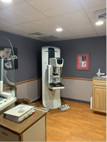 A mammogram is an image of the breast, done to detect breast cancer, most often in women over the age of 40. We are excited to announce the arrival of our NEW mammography machine! Starting in April, 2025 we will be offering diagnostic mammography services! LEARN MORE
CT Scan
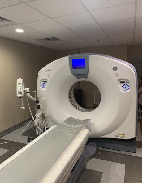
Computed Tomography, also known as CAT Scan, uses a limited beam of x- ray to obtain image data. This data is then interpreted by a computer to show cross sectional images of the body tissues and organs. Dense tissues, such as bones, appear white in the pictures. Less dense tissues, such as brain tissue or muscles, appear shades of gray and are air filled spaces, such as the bowel or lungs, appear black on the CT Scan.
Many CT Scans involve a prep of some sort, which will have been given to the patient by the practitioner's office or the Imaging Department. Contrast media is commonly used in the CT to opacify the GI tract and is usually taken orally andan hour or two before the scan. Another contrast media that contains iodine is injected intravenously during the scan. This makes the blood vessels and other structures more visable on the scan.IV contrast is often used to obtain images of the chest abdoman and pelvis and oral contrast is given for abdominal and/or pelvic CT Scans.
During the exam, the patient moves through the large opening of the scanner, the table moves up and slowly through and the tube inside it rotates, gathering the images, or data. This is then sent to the computer for interpretation. The exam itself is very quick. Here at Gordon Memorial Hospital we have a GE Optima 660 64-slice scanner.
Dexa Scans
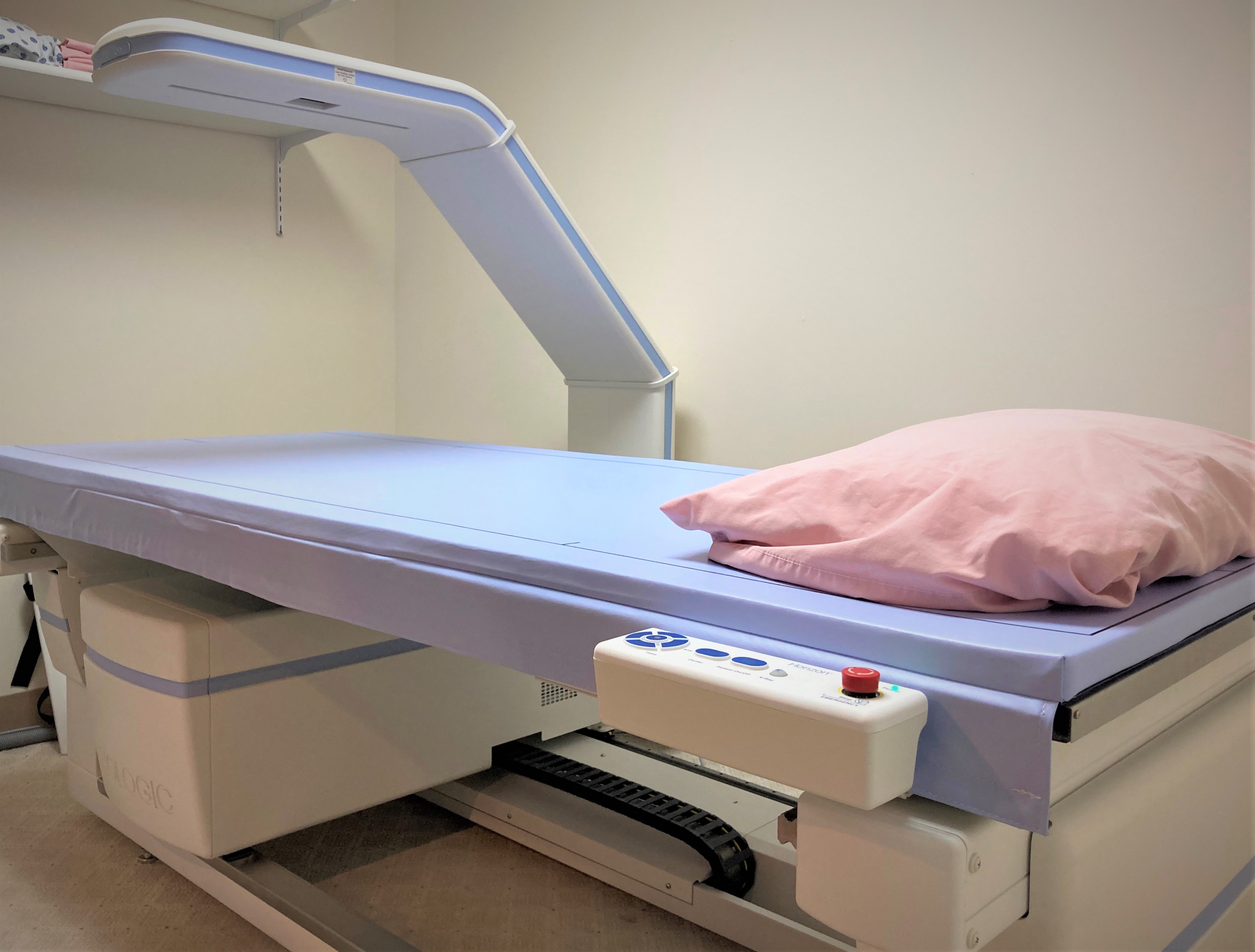
The DEXA Scan (Dual X-ray absorptiometry) is a widely used technique for measuring bone density to test for osteopenia or osteoprosis in men and women. A Dexa scan is a quick and painless procedure that measures bone loss in the spine and hips.
Osteoporosis is a gradual loss of calcium that causes bones to become thinner. This test can asses your risk for developing bone fractures and breaks. If the Dexa scan detects low bone density, your provider can help you develop a personalized plan to prevent further bone loss.
Our brand new Dexa Scanner is a wonderful asset that Gordon Memorial Hospital has been luckly enough to establish through the Overmass fondation. The Overmass fondation has contributed greatly to the health and welness of our small community and the surrounding areas.
Ultrasound
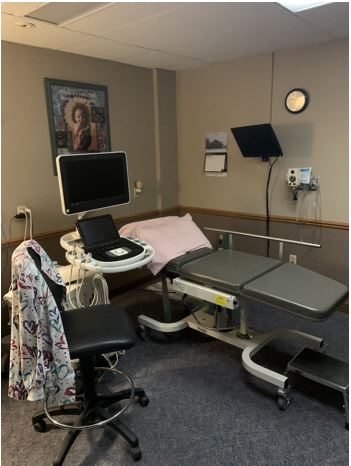
Ultrasound imaging, also called sonography is a method of obtaining images for inside the body by using high frequency sound waves. No radiation is used. Ultrasound is a useful way to examine many of the body's internal organs such as the liver, gallbladder, spleen, pancreas, kideys, bladder, ovaries, thyroid, and of course the anatomy of a fetus as it develops.
The preps for an ultrasound may vary and will be given to the patient by the ordering practitioner's office or the imaging dept. Most often for abdominal scans, the patient will have to NPO (nothing to eat or drink) for at least 6 hours and for pelvic scans will need a full bladder.
Vascular Ultrasounds (Doppler) Echocardiography
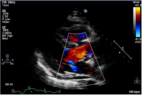
Dopplers & echocardiography are ultrasounds that examines blood flow. These exams are done by ultrasound technologists and the techs provided by Blackhills Ultrasound and the results are sent to the patients practitioner within a few days. Vascular ultrasounds are performed every other week on Wednesday.
 Magnetic Resonance Imaging (MRI) uses radiowaves and a strong magnetic field rather the x-rays to produce detailed images of body tissues and organs. The magnetic field 'excites' rather than 'relaxes' protons in the body, emitting radio signals. Those signals are processed by a computer to form an image.
 MRA (Magnetic Resonance Angiography) is also performed at Gordon Memorial Hospital. This exam provides detailed images of blood vessels with or without the use of contrast. The contrast is different than that used for CT Scans. The risk of an allergic reaction or kidney damage is very low. The amount given is based on the patients weight. Contrast may also be used for other MRI exams as requested (some neurologic studies always require contrast)
Due to strength of the magnet, all MRI patients are required to fill out a safety questionaireprior to their exam. Clothing should be free of all metal, jewelry, piercings, removable dental work, hairpins, etc will be removed.
The entire scan takes 45-60 minutes and results are sent the ordering practitioner within 48 hours of the exam.
MRI's are performed every Thursday, unless otherwise notified. MRI service is provided by Shared Medical Services.
